

Ndt Testing By X-Ray Crawler
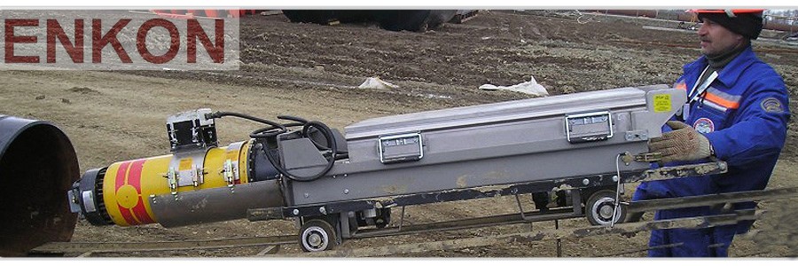
Automated X–ray systems for testing of weld joints use x–ray tubes or gamma–heads mounted on crawlers, which move through the pipe.
Crawlers allow the positioning of the radiation source precisely in the center of the pipe and can panoramically expose the weld to radiation .In this way, the image of the weld is displayed on the x–ray film in one exposure.
Panoramic radiographs, produced with x–ray tubes ,can easily be interpreted with the help of a weld defect catalogue.
- Crawlers ensure high–quality radiographs of girth weld joints during construction of mainline oil and gas pipelines with a range of pipe diameters from 135 mm to 1800 mm at temperatures down to –40oC
- The self–propelled trolley of the crawler has independent battery power to the drive units and the x–ray tube.
- The x–ray tube with constant potential gives smaller focus spot delivering higher density images and therefore renders to higher defect detection ability.
- The radiation parameters are stored in a special energy independent memory that is preserved even when the batteries are discharged.
- When the battery state reaches a pre–determined level, defined by the distance covered by the crawler, a recovery system automatically switches on, giving the command sequence for the crawler to exit the pipe.
- Crawlers are designed using highly integrated modules to facilitate assembly, make them more serviceable and more flexible to use.
- Isotopes or magnetic markers are placed on the external surfaces of the weld to be inspected for the exact positioning of the crawler.
Inspection through two walls is an alternative method for radiographic inspection of weld joints.
In this case it is not possible to have a panoramic shot of the pipe wall so the inspection has to be made in sections.
The x–ray film is placed on the external surface of the pipe section to be inspected and the x–ray source is placed against the pipe wall on the opposite side. This way, the section of the weld joint is radiographed through two walls.
The wall closest to the x–ray source is out of focus, but even then its shadow in the image can make the interpretation of the radiograph more difficult.
The productivity of this method is lower than when using an automated inspection system. Moreover this method requires a higher level of qualification from the personnel both to carry out the work and to interpret the results.
Results received from semi–automatic and annual measurements (ultrasonic, eddy current, dye penetrants, magnetic particle) are slightly more difficult to read and interpret and therefore require a more experienced operator.


